Posts

Pro.Rao Sripathi - Dentistry 2018 -Speaker

Image
Dr B.H. Sripathi Rao , Professor and Head of the Department of Oral and Maxillofacial Surgery is the present Principal/ Dean of Yenepoya Dental College, Yenepoya, Mangalore, Karnataka, India. He has wide experience in Orthognathic surgeries, Facial Trauma, Oral Cancer surgeries, Pathologies and Cleft Surgeries .He has been in maxillofacial Surgery teaching and training program for more than 35 years now with numerous publications and had been invited guest speaker to various international and national forums .He was also an executive member of the Dental Council Of India. ABSTRACT : Nasal Geometry – Changes after Maxillary Osteotomies and Corrective Measures The primary objective of all orthognathic surgeries inconjuction with proper occlusion is to improve facial esthetics A change in Nasal Geometry after Maxillary osteotomy is a common pertinent problem encountered by all maxillofacial surgeons. With changes in the facial skeleton following surgery, the facial drapes adapts to

Serap Titiz M.S - Speaker - Dentistry 2018

Image
Dr. Serap Titiz  Graduated from Hacettepe University Faculty of Dentistry in 2002.   She started his postgraduate education in 2012 at Ege University Faculty of Dentistry Department of Orthodontics and completed his PhD in 2012. She was appointed Assistant Professor in Orthodontics at Usak University Faculty of Dentistry since 2017. Abstract : A New Approach in Presurgical Nasoalveloar Molding Treatment in the Management of Cleft Lip and Palate Patients Objective: Many techniques have been used to align cleft alveolar segments and reduce cleft severity in cleft lip and palate patients The Grayson technique is a well-known technique that has undergone several modifications. The aim of this study was to present a new approach to PNAM for bilateral and unilateral cleft lip and palate patients. Material Method: In unilateral cleft lip and palate patients modified nostril retainers manufactured from soft acrylic were used instead of nasal stents for nasal molding w

Is Fluoride Safe ?

Image
Fluoride  is a natural mineral found throughout the earth’s crust and widely distributed in nature. Some foods and water supplies contain fluoride.Fluoride is found naturally in soil, water, and foods. It is also produced synthetically for use in drinking water, toothpaste, mouthwashes and various chemical products. Fluoride is often added to drinking water to help reduce tooth decay.Water authorities add fluoride to the  municipal water supply,  because studies have shown that adding it in areas where fluoride levels in the water are low can reduce the prevalence of tooth decay in the local population.Enamel, the outer layer of the crown of a tooth, is made of closely packed mineral crystals. Every day, minerals are lost and gained from inside the enamel crystals. Losing minerals is called demineralization. Gaining them back is called remineralization. Demineralization  begins with the bacteria in the plaque on your teeth. They feed on sugar and other carbohydrates in your mouth

DENTAL BRACES

Image
Not everyone is born with a  perfect teeth , some are lucky to be born with perfect teeth.Orthodontic treatment has now marked a revolutionary change in Dentistry, it helped to restore the smile with the help of Dental braces .Using this modern Dental technology it is now possible to bring back healthy and beautiful smile quicker. TYPES OF DENTAL BRACES Old-fashioned braces use elastic ties – which can cause painful friction – to hold the straightening wire in place.  They are made from a high-grade stainless steel and have metal brackets that are attached to each tooth using a type of cement. The brackets are linked to each other with a thin archwire, which puts pressure on the teeth to cause them to move slowly into the correct position.The ties are also prone to collecting plaque, which can be unpleasant. They are durable and strong. Ceramic Braces  : Made of Ceramics instead of Metal, they have clear or tooth colored brackets so they are less noticeable than metal brace

DENTAL RADIOGRAPHY- A Short Note

Image
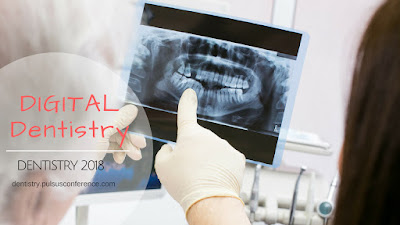
Radio graphs (X-rays) are necessary for the evaluation and diagnosis of many oral conditions and diseases. Dentists use radiographs for many reasons: to find hidden dental structures, malignant or benign masses, bone loss, and cavities. They are diagnostic, but they can also be preventative, by helping a dentist diagnose potential oral care issues in a patient’s mouth before they become a major problem. An x-ray is a type of energy that passes through soft tissues and is absorbed by dense tissue. Teeth and bone are very dense, so they absorb X-rays, while X-rays pass more easily through gums and cheeks.X-rays are divided into two main categories, intraoral and extraoral. Intraoral is an X-ray that is taken inside the mouth. An extraoral X-ray is taken outside of the mouth. Intraoral X-rays are the most common type of radiograph taken in dentistry. The benefits of X-rays are well known: They help dentists diagnose common problems, such as cavities, gum disease and some types

TOOTH DISCOLORATION

Image
Tooth discoloration can be the cause of great embarrassment and can make people feel self conscious about smiling .Teeth Stains, also commonly referred to as  Teeth Discoloration , is caused by substances sticking onto the surface of your tooth or gathering within and contaminating the enamel of your tooth.Nevertheless, important lifestyle factors also play a large part in tooth discoloration and it is important to recognize these. Symptoms include stains on the enamel. They can range from white streaks to yellow tints or brown spots and pits. If the enamel has worn away, and dentin is showing through, you may notice a yellow tint. Some food habits and lifestyle that can cause tooth discoloration:  Food and drink.  Coffee, tea, carbonated beverages and red wine can discolor teeth. Even certain fruits and vegetables can cause stain Poor dental hygiene .  Inadequate brushing, flossing, and rinsing with an antiseptic mouthwash to remove plaque and stain-producing subst

GINGIVITIS

Image
Gingivitis  means inflammation of the gums, commonly occurs because a film of plaque, or bacteria, accumulates on the teeth. Gingivitis is a  non-destructive  type of periodontal disease, but untreated gingivitis can progress to periodontitis in which the inflammation of the gums results in tissue destruction and bone resorption around the teeth.  Periodontitis  can ultimately lead to tooth loss. Signs of gingivitis include red and puffy gums, that bleed easily when the person brushes their teeth, Bad breath and Swollen gums. The most common cause of gingivitis is the accumulation of bacterial plaque between and around the teeth. The plaque triggers an immune response, which, in turn, can eventually lead to the destruction of gingival, or gum, tissue. It may also, eventually, lead to further complications, including the loss of teeth. Dental plaque  is a biofilm that accumulates naturally on the teeth. It is usually formed by colonizing bacteria that are trying to stic