DENTAL RADIOGRAPHY- A Short Note
They are diagnostic, but they can also be preventative, by helping a dentist diagnose potential oral care issues in a patient’s mouth before they become a major problem. An x-ray is a type of energy that passes through soft tissues and is absorbed by dense tissue. Teeth and bone are very dense, so they absorb X-rays, while X-rays pass more easily through gums and cheeks.X-rays are divided into two main categories, intraoral and extraoral. Intraoral is an X-ray that is taken inside the mouth. An extraoral X-ray is taken outside of the mouth. Intraoral X-rays are the most common type of radiograph taken in dentistry.
The benefits of X-rays are well known: They help dentists diagnose common problems, such as cavities, gum disease and some types of infections. Radiographs allow dentists to see inside a tooth and beneath the gums to assess the health of the bone and supporting tissues that hold teeth in place.The dosage of X-ray radiation received by a dental patient is typically small (around 0.150 mSv for a full mouth series.Dentists use X-rays to help diagnose damage and disease that is not visible during a clinical dental examination. How often X-rays should be taken depends on specific factors such as an individual’s current oral health, age, risk for disease and any signs or symptoms of oral disease.
The periapical (PA) view is taken of both anterior and posterior teeth. The objective of this type of view is to capture the tip of the root on the film. This is often helpful in determining the cause of pain in a specific tooth, because it allows a dentist to visualize the tooth as well as the surrounding bone in their entirety.
The bitewing view is taken to visualize the crowns of the posterior teeth and the height of the alveolar bone in relation to the cementoenamel junctions, which are the demarcation lines on the teeth which separate tooth crown from tooth root. Routine bitewing radiographs are commonly used to examine for interdental caries and recurrent caries under existing restorations.
Dental X-rays are very safe and expose you or your child to a minimal amount of radiation. When all standard safety precautions are taken, today's X-ray equipment is able to eliminate unnecessary radiation and allows the dentist to focus the X-ray beam on a specific part of the mouth. High-speed film enables the dentist to reduce the amount of radiation the patient receives. A lead body apron covers the body from the neck to the knees and protects the body from stray radiation.
Bite-wing x-rays show details of the upper and lower teeth in one area of the mouth. Each bite-wing shows a tooth from its crown (the exposed surface) to the level of the supporting bone.
Periapical x-rays show the whole tooth — from the crown, to beyond the root where the tooth attaches into the jaw. Each periapical x-ray shows all teeth in one portion of either the upper or lower jaw.
Occlusal x-rays track the development and placement of an entire arch of teeth in either the upper or lower jaw.
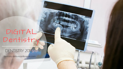
Extraoral x-rays are used to detect dental problems in the jaw and skull. There are several types of extraoral x-rays.
Cone Beam CT is a type of x-ray that creates 3-D images of dental structures, soft tissue, nerves, and bone. It helps guide tooth implant placement and evaluates cysts and tumors in the mouth and face. It also can detect problems in the gums, roots of teeth, and jaws. Cone beam CT is similar to regular dental CT in some ways.
Digital imaging is a 2-D type of dental imaging that allows images to be sent directly to a computer. The images can be viewed on screen, stored, or printed out in a matter of seconds. Digital imaging has several other advantages compared with traditional x-rays.
MRI imaging is an imaging method that takes a 3-D view of the oral cavity including jaw and teeth.


Yeah, I agree with this post, Efficiency in capturing X-rays also translates to time saved. Less time spent developing film X-rays and taking patients through their treatment plans means more time was treating patients. Over weeks and months, the minutes wasted developing film turn to hours.
ReplyDeleteABS - www.atlantabasedsystems.com
Nice information about Dental x-rays test
ReplyDelete