Pro.Rao Sripathi - Dentistry 2018 -Speaker
 Dr B.H. Sripathi Rao, Professor and Head of the Department of Oral and Maxillofacial Surgery is the present Principal/ Dean of Yenepoya Dental College, Yenepoya, Mangalore, Karnataka, India. He has wide experience in Orthognathic surgeries, Facial Trauma, Oral Cancer surgeries, Pathologies and Cleft Surgeries .He has been in maxillofacial Surgery teaching and training program for more than 35 years now with numerous publications and had been invited guest speaker to various international and national forums .He was also an executive member of the Dental Council Of India.
ABSTRACT :
Nasal Geometry – Changes after Maxillary Osteotomies and Corrective Measures
The primary objective of all orthognathic surgeries inconjuction with proper occlusion is to improve facial esthetics A change in Nasal Geometry after Maxillary osteotomy is a common pertinent problem encountered by all maxillofacial surgeons. With changes in the facial skeleton following surgery, the facial drapes adapts to the new changes in the supporting skeleton in a manner that is not always uniform. This is because the soft tissue drapes varies in thickness and tone from region to region and from individual to individual.
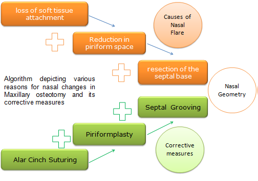
Firmly attached soft tissues exhibit more consistent changes while unattached tissues show less consistent changes due to their resiliency and elasticity.
The type and magnitude of the Neo- Skeletal position creates nasolabial changes which is a challenging clinical situation to handle as it is not always predictable. The changes can range from elevation or depression of the nasal tip, increase in the nasolabial angle and increase in the alar base width.
In our study, we analyzed the efficacy of the adjunctive procedures like Alar cinch Suturing, Piriformplasty and septal grooving alone and in combination after Vertical repositioning of the Maxilla. This paper discusses the reasons of changes in nasal geometry after maxillary osteotomy and draws a consensus in protocol building for corrective measures in nasal changes after maxillary osteotomies, particularly after superior repositioning.
Recent Publications
1. Warner JP, Chauhan N, Adamson PA. Alar soft-tissue techniques in rhinoplasty: algorithmic approach, quantifiable guidelines, and scar outcomes from a single surgeon experience. Arch Facial Plast Surg. 2010. May-Jun;12(3):149–58. 10.1001/archfacial.2010.30
2. Manisali M, Khamashta-Ledezma L. Rhinoplasty and nasal changes in relation to orthognathic surgery. Oxford: Wiley- Blackwell; 2017. pp. 724–38.
3. Liu X, Zhu S, Hu J. Modified versus classic alar base sutures after LeFort I osteotomy: a systematic review. Oral Surg Oral Med Oral Pathol Oral Radiol. 2014. January;117(1):37–44. 10.1016/j.oooo.2013.09.002


Comments
Post a Comment